Sydwest Eye Specialists is one of the renowned ophthalmology centres in the city of Cabramatta, NSW. Sydwest Eye Specialists is featured with well equipped state-of-the-art diagnostic & treatment equipments that help the doctors in diagnosing and treating the eye disorders effectively and also in giving the best possible medical service to the patients. Some of the most advanced equipments available at Sydwest Eye Specialists include:
Sydwest Eye Specialists is the only practice in Sydney`s South West that has this technology.
The advanced technology of optomap ultra-wide digital retinal imaging system helps your doctor to detect, diagnose and manage early signs of retinal diseases or even signs of some systemic diseases more efficiently and effectively. The digital imaging system views up to 80% or 200 degrees of your retina in one single capture compared to 10-15% or 45 degrees with the traditional methods. This diagnostic tool allows your doctor to view most of the retina at one single time of retinal capture. The procedure is performed in seconds, doesn’t require retinal dilation and is a non-painful procedure. Optomap will also allow your eye care professional to have a digital record of your retinal health which can be compared for the changes over time.
Cirrus OCT – It is one of the most advanced optical coherence tomography (OCT) system used to view the retina, back layer of the eye, including the optic nerve and the macula. It is manufactured by Carl Zeiss Meditec, an integrated medical technology company. This allows for early detection of retinal disease accurately and objectively, designing of treatment protocol and also optimises evaluation of pre- and post- therapy changes. This new high-performance OCT instrument delivers exquisite high-definition images of ocular structures. It is used for the management of glaucoma and retinal disease, retina assessment for cataract surgery, and anterior segment imaging for corneal disease. The Cirrus OCT is organized with the most advanced applications, such as advanced RPE (retinal pigment epithelial) analysis and ganglion cell analysis, retinal tracking, and macular thickness and change analysis. The new Cirrus OCT allows more practitioners to offer patients a high level of care.
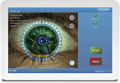
New Image Guided System
Precise Surgical Planning and Procedures
The new VERION™ Image Guided System is designed to offer improved precision, consistency and control in cataract refractive surgery.
Improved Patient outcomes:
- Minimize data recording errors
- Less patient discomfort by shorter procedure time
- Increase toric and multifocal IOL confidence
- More consistent and reliable surgical outcomes
- Optimize precision and visual results
Humphrey Visual Field Analyzer – It is the accepted standard of care in glaucoma diagnosis and management. This is the premier automated visual field perimeter to measure visual field of the eye. Visual field test measures the capacity of your eye in ‘side vision’. Visual field may be impaired in the diagnosis of conditions which affect the visual field such as glaucoma, macular degeneration, retinal detachment, diabetic retinopathy, certain neurological conditions and other conditions affecting the optic nerve of the eye.
Zeiss Visucam fundus camera – It is a digital camera from Zeiss that provides photographs of the inner surface of the eye including fundus, macula, optic disc and retina. The fundus cameras are used for diagnosing the condition, monitoring the progression of a disease, and in screening procedures. Zeiss Visucam Fundus Camera delivers two-field angles the standard 45° and the smaller 25° field for excellent photos of the optic disc and macula. It is valuable in the diagnosis of glaucoma, diabetic retinopathy and other retinal diseases. It provides both red colour and red free colour taking modes; red free colour mode is used to visualize the blood vessels on the retina after angiography.
IOL Master – It is a non-contact optical instrument to measure the eye length and surface curvature of the eye accurately and without pain. These two parameters are required essentially before cataract surgery for calculation and selection of the right power of the artificial lens to be used for implantation. It uses partial coherence interferometry technology utilising laser for the measurement and is the best technology available for the purpose. It gives five times more accurate measurements than A-scan ultrasonography. As the IOL master is non-contact instrument there are no chances of contamination and also anaesthesia is not required.
Atlas Cornea Topographer – It is a latest device used to create topographic map of the cornea which is similar to a contour map of the surface of the land. Atlas Cornea Topographer combines the highly sophisticated software programs with most advanced imaging and analysis technology. It is used in planning and evaluation of LASIK surgery and for the diagnosis of keratoconus. It is also used to assess the appropriate fit of the contact lenses especially for people suffering from keratoconus, in diagnosis and management of corneal conditions and procedures such as corneal transplants, corneal opacities, corneal deformities, post-operative cataract extraction with acquired astigmatism.
Argon Laser – Laser is a light energy used for treating various eye conditions and also skin conditions. Laser devices emit the radiation which when focused using microscope destroys the tissues. Argon laser is the laser device where argon is used as amplifying medium to intensify the energy. Laser treatment is more precise, safe and effective and also cost-effective. Argon laser can pass through the fluids inside the eye without damaging the surrounding tissues and therefore used in treatment of diabetic retinopathy. Other applications include argon laser trabeculoplasty, argon laser treatment for vascular diseases, retinal breaks, retinal degeneration and selected eye tumors.
Autorefractor – Autorefractor is one of the advanced computerised instruments used for eye examination to assess the refractive error and aid in prescribing glasses or contact lenses. This simple instrument evaluates the refractive error of a patient by measuring how the light is changed after entering into eye. The procedure is painless, quick and does not require the patient’s feedback to assess the refractive power.
Zeiss forum integration software – The Zeiss FORUM integration software centralizes and stores data from the ophthalmic diagnostic instruments which enables your doctor to access data from any instrument. This software increases flexibility by sharing raw data of multiple diagnostic instruments through a central database. When this data sharing is enabled, independent functioning of the patient exams and their analysis occurs without the need of individual diagnostic instruments. The archiving ability of the software keeps the data safe, maintains consistency across all diagnostic instruments and systems, and is useful to integrate with multiple CIRRUS OCTs or visual field analyzers. It provides immediate access to all patient data from wherever and whenever you want, thereby increasing the workflow efficiency and management of data from multiple patient visits.